






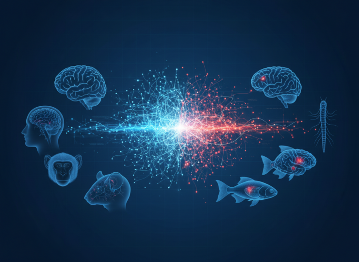
General anaesthesia can make a human, a monkey, a fish or even a tiny nematode stop responding to the outside world. The drugs, nervous systems and measurement methods can differ dramatically, yet a new cross-species analysis suggests that the resulting brain state converges on a surprisingly similar dynamical pattern.
Research published in Nature Neuroscience on 29 September 2026 found that anaesthesia consistently shortened the intrinsic timescales of neural activity and weakened coordination between different parts of the nervous system. Across six species spanning roughly 700 million years of evolution, the common endpoint was what the researchers describe as spatiotemporal isolation of local neural activity.
A search for what different anaesthetics have in common
Anaesthetic drugs do not all work through the same molecular targets. Researchers have also traditionally studied anaesthesia in individual species and often selected a relatively small number of familiar signal properties, such as spectral power or entropy.
Andrea Luppi and colleagues took a broader approach. They assembled multiscale neural recordings from humans, macaques, marmosets, mice, larval zebrafish and Caenorhabditis elegans nematodes, then compared awake and anaesthetised states across several different drugs and recording techniques.
The mammalian datasets primarily used functional magnetic resonance imaging, while zebrafish and nematodes were studied with calcium imaging. The collection included 15 awake-versus-anaesthesia or re-awakening contrasts and seven anaesthetic regimes, allowing the team to look for effects that survived large differences in biology, drug action and measurement scale.
The human dataset included 15 healthy participants with usable resting-state fMRI data at baseline, during deep sevoflurane anaesthesia and during recovery. Other datasets included five macaques exposed to sevoflurane, propofol or ketamine, four marmosets studied with three anaesthetics, and 43 mice across awake, halothane and medetomidine-isoflurane conditions.
The analysis extended beyond mammals. Seven larval zebrafish contributed neuronal calcium imaging during wakefulness and tricaine anaesthesia, while ten nematodes were studied during exposure to isoflurane.
More than 6,000 features narrowed to a common signature
Rather than deciding in advance which signal properties mattered most, the researchers extracted more than 6,000 time-series features from every brain region or neuron. These covered properties such as autocorrelation, periodicity, stationarity, complexity, distribution shape and self-similarity.
The team then imposed an unusually strict consistency test. A feature was retained only if its direction of change under anaesthesia was the same across every relevant species and contrast. One contradictory result in one dataset was enough to remove it from the common set.
That screen identified 485 features that changed consistently across regions, anaesthetics and species. Under a simple sign-based chance model, a feature would have only a 1 in 16,384 probability of showing the same direction across all 15 contrasts by chance.
The similarity was not merely a matter of all features moving in the same direction. The full dynamical signatures were strongly correlated across different species and drugs, with a mean correlation of 0.86. A permutation test indicated that this agreement was substantially greater than expected simply from selecting features with matching signs, with p below 0.0001.
Anaesthetised activity became faster locally and less coordinated globally
The shared signature pointed to shorter intrinsic neural timescales. In practical terms, activity under anaesthesia retained less influence from its recent past and decayed more rapidly, reducing the temporal continuity that characterises the awake state.
At the same time, communication across the nervous system became less coordinated. Anaesthesia reduced synchronous cofluctuation between regions and made their dynamical profiles less similar to one another.
The combination is important because a brain can contain locally active regions without those regions operating as a coordinated whole. The authors interpret the cross-species pattern as a form of spatiotemporal isolation: local activity becomes more self-contained in time and less integrated with activity elsewhere.
This offers a possible bridge between neural signals and the behavioural hallmark of general anaesthesia. An organism may stop responding to its environment not because neural activity simply shuts down, but because the temporal persistence and large-scale coordination needed to integrate information are disrupted.
Deep-brain stimulation reversed the signature
One of the strongest tests came from a macaque dataset in which propofol remained in the bloodstream while researchers stimulated different thalamic sites. Stimulation of the centromedian thalamus restored behavioural responsiveness, while stimulation of a control thalamic site did not.
When the animals re-awakened during effective stimulation, their neural dynamics shifted in the opposite direction to anaesthesia. Intrinsic timescales lengthened and the broader dynamical profile moved back toward the awake state.
Across 82 brain regions, the degree to which intrinsic timescales increased during successful centromedian stimulation was strongly related to the regions’ awake-state values. The reported Spearman correlations were -0.95 when effective stimulation was compared with propofol without stimulation and -0.90 when compared with ineffective control-site stimulation, both with p below 0.001.
That reversal strengthens the argument that the identified dynamics track responsiveness rather than merely the physical presence of an anaesthetic drug. It also provides a rare example of the same neural signature moving in both directions as behavioural state changes.
The pattern also connected to genes and synaptic biology
The researchers next asked whether the conserved dynamics could be related to conserved molecular organisation. They compared cortical patterns of 23 orthologous brain-related genes across humans, macaques, marmosets and mice, including genes involved in glutamate, GABA and other neurotransmitter systems.
A multivariate analysis identified a statistically significant relationship between gene-expression patterns and anaesthetic-induced changes in neural dynamics. This does not prove that those genes cause the observed state, but it connects the large-scale signal pattern to biological systems that are conserved across species.
The team also tested a previously developed biophysical model in which anaesthesia prolongs inhibitory postsynaptic currents. Remarkably, the model reproduced the direction of change for 461 of the 485 empirically consistent features, or about 95%, significantly more than expected by chance.
That modelling result offers a plausible route from molecular and synaptic effects to the large-scale dynamics visible in whole-brain recordings. It remains a mechanistic model rather than direct proof that one synaptic process explains every anaesthetic or every species.
What the study can and cannot tell us
The breadth of the dataset is a major strength, but it also creates limitations. The species were recorded with different technologies, spatial resolutions, preprocessing pipelines, drugs and doses. Sample sizes were also small in several animal datasets, including five macaques, four marmosets and seven zebrafish.
The analysis deliberately focused on what was shared across species and anaesthetics. It therefore does not imply that every anaesthetic produces an identical brain state, nor that species-specific differences are unimportant. The authors observed differences alongside the common signature and identify them as a target for future work.
Loss of behavioural responsiveness is also an imperfect proxy for consciousness. The study is strongest as an account of the neural dynamics accompanying anaesthetic-induced disconnection from the environment, rather than a complete explanation of subjective awareness.
Even with those boundaries, the cross-species convergence is striking. A human cortex, a fish nervous system and a nematode neural network are separated by enormous evolutionary and anatomical distances, yet anaesthesia pushed their activity toward a recognisably shared dynamical regime.
The result suggests that the ability to remain responsive may depend on a very old principle of nervous-system organisation: neural activity must persist for long enough and coordinate broadly enough for information to be integrated across space and time. Anaesthesia appears to disrupt both properties together.
Source Information
Study Title: Comprehensive profiling of brain dynamics during anesthesia across phylogeny
Authors: Andrea I. Luppi, Lynn Uhrig, Jordy Tasserie, Golia Shafiei, Antoine Légaré, Kanako Muta, Junichi Hata, Hideyuki Okano, Daniel Golkowski, Andreas Ranft, Rudiger Ilg, Denis Jordan, Silvia Gini, Zhen-Qi Liu, Yohan Yee, Pablo Castro, Camilo M. Signorelli, Rodrigo Cofre, Alain Destexhe, David K. Menon, Emmanuel A. Stamatakis, Patrick Desrosiers, Paul De Koninck, Christopher W. Connor, Alessandro Gozzi, Ben D. Fulcher, Bechir Jarraya, Bratislav Misic and colleagues
Journal: Nature Neuroscience
Year: 2026
DOI: 10.1038/s41593-026-02460-4