







Cells that stop dividing are not always useless. Temporary cellular senescence can help with processes such as wound repair, but senescent cells that persist with age can release inflammatory signals and contribute to tissue dysfunction. A new study identifies a cellular recycling pathway as one reason these cells become harder for the immune system to remove as organisms grow older.
Published in Nature Aging on 5 October 2026, the research focuses on chaperone-mediated autophagy, or CMA, a selective form of protein degradation inside lysosomes. The experiments show that age-related CMA decline changes the signals released by senescent cells and weakens the ability of macrophages to engulf and remove them. In aged mice, pharmacologically boosting CMA reduced senescent cell burden across multiple tissues and improved measures of fibrosis.
The findings provide a mechanistic explanation for a long-standing problem in aging biology: why senescent cells that are normally cleared by the immune system increasingly accumulate in older tissues. They also identify CMA as a potential therapeutic target, although the evidence remains preclinical and does not establish that activating the pathway will safely slow aging or treat age-related disease in people.
Why senescent cells become a problem with age
Senescence is a stress response in which damaged or otherwise challenged cells enter a durable cell-cycle arrest. In younger organisms, this state can be transient because immune cells, including macrophages, natural killer cells and T cells, recognize and remove senescent cells. With age, however, senescent cells increasingly escape immune clearance.
That persistence matters because senescent cells secrete a complex mixture of proteins and metabolites known as the senescence-associated secretory phenotype, or SASP. These signals can influence inflammation, neighboring cells and immune responses. Accumulation of senescent cells has therefore become an important target in research on age-associated disease.
The new study connects this process with another hallmark of aging: declining autophagy. CMA differs from bulk forms of autophagy because it selectively identifies proteins carrying particular targeting motifs and transports them into lysosomes for degradation. CMA activity declines with age in many tissues and has previously been linked to neurodegeneration, metabolic dysfunction and atherosclerosis.
Old cells failed to increase CMA when they became senescent
The researchers began with primary mouse ear fibroblasts from young, four-month-old mice and aged, 23-month-old mice. Using a fluorescent reporter that allows CMA activity to be monitored, they found that young cells increased CMA after senescence was induced. Aged cells started with lower CMA activity and failed to mount the same increase.
Importantly, simply blocking CMA did not fully turn young cells into conventional senescent cells. Instead, CMA-deficient cells developed a collection of proteomic, metabolic and secretory characteristics resembling those seen in aged senescent cells. This distinction suggests that declining CMA does not merely trigger senescence. It changes the type of senescent state that develops and, critically, the signals those cells release.
The secretome from CMA-deficient senescent cells had stronger pro-senescence effects on neighboring cells. It also suppressed CMA activity in macrophages, creating the possibility of a feedback loop in which aging cells make immune clearance progressively less effective.
Macrophages lost some of their ability to clear senescent cells
Macrophages are professional engulfing cells that remove damaged cells and cellular debris. The researchers found significantly reduced CMA activity in bone marrow-derived macrophages from aged mice of both sexes. Exposure to secretions from senescent fibroblasts also reduced CMA in young macrophages, with an even stronger inhibitory effect from CMA-deficient cells.
To test whether this loss was functionally important, the team used mice in which CMA was selectively impaired in macrophages. Macrophages lacking the CMA receptor LAMP2A showed reduced phagocytic and efferocytic activity and were less effective at clearing senescent fibroblasts in co-culture. The study traced part of this defect to impaired internalisation of inhibitory receptors involved in the cellular “do not eat me” signalling system.
The consequences extended beyond a dish. In an acute wound-healing experiment using 12-month-old mice, animals with macrophage-specific CMA deficiency had a significantly larger remaining wound area 15 days after injury, delayed closure and more p21-positive cells in the damaged tissue despite greater macrophage abundance. This indicates that having macrophages present is not enough if their clearance machinery is compromised.
Five months of CMA activation reduced senescence markers in aged mice
The most translational part of the study asked whether restoring CMA could reverse some of these age-associated changes. Researchers gave 18-month-old mice a previously developed small-molecule CMA activator orally for five months, at 30 milligrams per kilogram of body weight per day. At the end of treatment, the mice were approximately 23 months old and were compared with age-matched vehicle-treated animals and young six-month-old mice.
CMA activation prevented the age-related increase in cells positive for senescence-associated beta-galactosidase in gonadal white adipose tissue, liver and lung in both sexes. It also reduced the age-associated expression of senescence-related markers including p21, TNF, IL-1 alpha and p16 across tissues. Histological analysis showed reductions in fibrosis, particularly in white adipose tissue, while multivariable analysis placed treated old animals closer to the young group across the measured senescence markers.
The intervention also improved macrophage function. A 48-hour treatment of macrophages isolated from 23-month-old mice restored their phagocytic activity to levels comparable with macrophages from six-month-old animals.
The pathway was also tested in experimental lung fibrosis
Because persistent senescence is implicated in pulmonary fibrosis, the researchers also examined CMA activation in an experimental mouse model of lung fibrosis. Systemic activation reduced senescent cell burden and attenuated disease severity. The intervention broadly changed the local immune environment, including macrophage, CD4-positive T-cell and neutrophil responses.
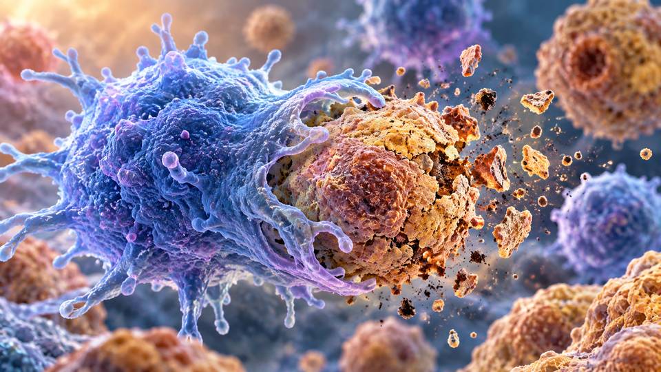
Together with the aging experiments, this supports the authors’ proposal that CMA has a dual role. It shapes the biology of the senescent cells themselves while also maintaining the immune cells responsible for removing them. When CMA declines on both sides of that interaction, senescent cells can become more difficult to clear and their secretions can further suppress macrophage function.
A feedback loop may help explain senescent cell accumulation
The study therefore points to a self-reinforcing mechanism. Aging reduces CMA in cells entering senescence. Those cells develop an altered SASP with stronger pro-senescence and immune-suppressing effects. Their secretions reduce CMA in macrophages, which then become worse at engulfing senescent cells. The surviving senescent cells can continue releasing signals that sustain the cycle.
This model is significant because it connects several biological features of aging that are often studied separately: loss of protein quality control, impaired autophagy, cellular senescence, immune dysfunction and fibrosis. Rather than representing independent failures, they may reinforce one another through shared molecular pathways.
What the findings do not yet show
The results are promising but should not be interpreted as evidence for an anti-aging drug in humans. Much of the mechanistic work used mouse fibroblasts and genetically modified mice. Senescence was commonly induced with palbociclib, although the researchers used additional senescence triggers in selected experiments and generally observed comparable effects.
The macrophage-specific genetic model also relies on LysM-Cre, which can affect other myeloid cells such as monocytes and granulocytes. Experiments using isolated macrophages support a direct macrophage role, but other immune cells may contribute to the effects observed in living animals.
Human evidence was limited. The researchers included lung data relevant to idiopathic pulmonary fibrosis, but these observations were correlative. CMA cannot yet be directly assessed in people in the same way it can in the experimental models used here. Long-term safety is another central question because senescence and autophagy have normal physiological roles, meaning that manipulating either pathway could have effects beyond the intended target.
Why the research matters
Strategies for dealing with senescent cells have often focused on killing them directly or suppressing the inflammatory signals they release. This research suggests another possibility: improving the body’s own ability to resolve senescence by restoring a cellular maintenance pathway that supports both senescent-cell biology and immune clearance.
That distinction could matter for future therapeutics. Rather than treating all senescent cells as identical targets, interventions might eventually aim to restore the processes that allow beneficial, temporary senescence to resolve while limiting the persistent form associated with aging and disease.
For now, the strongest conclusion is mechanistic. Declining chaperone-mediated autophagy appears capable of turning senescence and immune clearance into a damaging feedback loop in aged mice, and experimentally restoring that pathway was sufficient to reduce several markers of senescent-cell accumulation. Whether the same approach can be translated safely to humans remains an open question.
Source Information
Study: Sereda, R., Lindenau, K., Diaz, A. et al. “Decline of chaperone-mediated autophagy in aging impairs macrophage clearance of senescent cells.” Nature Aging (2026).
Published: 5 October 2026
DOI: 10.1038/s43587-026-01240-w