






Stress is often discussed as if it affects the mind and the body through separate pathways.
New research suggests that the connection can be much more direct.
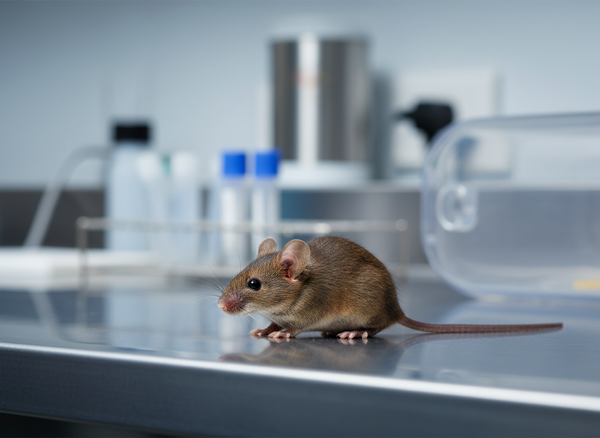
A study published in Nature Communications on 24 September 2026 found that chronic stress promoted the progression of hepatocellular carcinoma in male mice through a nerve circuit linking the brain and liver.
The pathway began in the hypothalamus, travelled through the vagus nerve and altered chemical signalling inside liver tumours.
The researchers also identified a feedback loop in which inflammatory signals produced around the tumour travelled back to the brain and intensified depression-like behaviour in the mice.
When parts of this circuit were interrupted, tumour progression slowed and the behavioural effects of chronic stress were reduced.
The findings provide a detailed biological mechanism linking chronic stress, the nervous system, immune signalling and cancer progression in mice.
They do not show that stress causes liver cancer in people.
The study focused on hepatocellular carcinoma
Hepatocellular carcinoma is the most common form of primary liver cancer.
Its development is influenced by factors such as chronic viral hepatitis, alcohol-related liver disease, metabolic dysfunction and cirrhosis.
Once a tumour is established, however, its progression can also be influenced by the surrounding immune system, blood supply, hormones and nerve signals.
The new study examined whether chronic stress could alter this tumour environment through communication between the brain and liver.
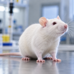
The researchers used male mouse models of hepatocellular carcinoma and exposed animals to chronic stress conditions while monitoring tumour growth, brain activity, immune signalling and behaviour.
They also used molecular and cellular experiments to identify the signals that connected these different systems.
Chronic stress activated a specific pathway from the brain to the liver
The first part of the pathway began in the paraventricular nucleus of the hypothalamus.
The hypothalamus helps regulate stress responses, hormones, autonomic functions and many other processes needed to maintain internal balance.
Under chronic stress, glutamatergic neurons in this region became more active.
These neurons influenced the dorsal motor nucleus of the vagus, a brainstem region that sends parasympathetic signals to internal organs through the vagus nerve.
The result was increased cholinergic signalling from the brain to the liver.
This created a functional pathway running from the hypothalamus to the vagus nerve and then to the tumour-bearing liver.
Stress increased acetylcholine inside the tumour
A key chemical in this pathway was acetylcholine.
Acetylcholine is a neurotransmitter used widely throughout the nervous system.
In the stressed mice, activation of the brain to liver circuit increased acetylcholine within the tumour environment.
This was important because some tumour epithelial cells expressed a receptor called CHRNA9, which can respond to acetylcholine.
The researchers found that acetylcholine promoted tumour-cell proliferation through this receptor.
This means the nervous system was not simply responding to the tumour.
In the mouse models, neural signalling directly altered conditions that supported tumour growth.
Single-cell analysis revealed which tumour cells responded
To examine the tumour environment in greater detail, the researchers used single-cell transcriptomics.
This method measures gene activity in individual cells rather than averaging signals across an entire tissue sample.
It can therefore reveal distinct cell populations that would otherwise be hidden inside a complex tumour.
The analysis identified a population of epithelial tumour cells expressing Chrna9, the gene encoding the CHRNA9 receptor.
These cells were particularly responsive to acetylcholine.
The finding helped connect the increased nerve signal to a specific molecular response inside the tumour.
Tumour cells then interacted with macrophages
The mechanism did not stop with the tumour cells themselves.
The acetylcholine-responsive epithelial cells interacted with macrophages, immune cells that can play very different roles depending on their local environment.
Some macrophages attack abnormal cells and support anti-tumour immunity.
Others can become part of a tumour-supporting environment and release signals that promote growth, blood-vessel formation or immune suppression.
In the stressed mice, communication between epithelial cells and macrophages involved COL1A1 and COL1A2, the CD44 receptor and STAT3 signalling.
This interaction increased production of the inflammatory chemokine CXCL1.
CXCL1 helped turn the pathway into a feedback loop
CXCL1 was not limited to the tumour environment.
The researchers found that circulating CXCL1 could cross the blood-brain barrier and influence the hypothalamus.
Once in the brain, it activated glutamatergic neurons in the same hypothalamic region involved in the original stress pathway.
This created a feedback loop.
Chronic stress increased neural signalling to the liver. The altered tumour environment then generated inflammatory signals that returned to the brain and reinforced the neural response.
The mice also displayed stronger depression-like behaviours as this loop became established.
In animal studies, the term depression-like behaviour refers to measurable behavioural changes used as experimental proxies. It should not be treated as identical to a clinical diagnosis of depression in humans.
The researchers disrupted the circuit in several different ways
A major strength of the study was that the researchers did not rely only on observing associations.
They interfered with different parts of the proposed pathway and examined what happened next.
Silencing the hypothalamic vagal circuit reduced tumour progression.
Knocking down Chrna9 specifically in tumour cells also restrained tumour growth.
Neutralising CXCL1 systemically produced another reduction in tumour progression.
These interventions also alleviated the depression-like behavioural changes observed in the stressed mice.
Because several different interventions affected the same overall pattern, the evidence for the proposed pathway is stronger than it would be if the study had identified only one correlation.
The pathway linked the nervous and immune systems
The study illustrates how difficult it can be to separate the nervous system from the immune system when studying disease.
Stress begins with changes in brain activity, but those changes can alter autonomic nerves, hormone release and immune function.
Tumours also communicate with surrounding immune cells and release molecules into the circulation.
Those circulating molecules can then affect distant organs, including the brain.
The new work places these processes into one connected mechanism in male mice with hepatocellular carcinoma.
The brain influenced the tumour, and the tumour environment fed signals back to the brain.
Other forms of chronic physiological stress showed a similar association
The researchers also examined systemic inflammation and neuropathic pain.
These conditions were associated with hepatocellular carcinoma progression in the male mice as well.
This suggests that the broader connection may not be limited to one particular experimental stressor.
However, the detailed mechanistic work in the paper focused on the chronic stress pathway.
The study therefore should not be interpreted as showing that all forms of pain, inflammation or psychological distress influence cancer through exactly the same mechanism.
The findings do not show that stress causes liver cancer in humans
This distinction is essential.
The experiments examined cancer progression in male mice that already had hepatocellular carcinoma models.
The study did not show that chronic stress creates liver cancer in otherwise healthy animals or people.
It also did not test whether ordinary life stress in humans produces the same biological pathway or changes survival among patients with liver cancer.
Human cancer is influenced by many interacting factors, including tumour genetics, liver function, treatment, age, metabolism, infections and underlying liver disease.
The mouse mechanism is therefore a starting point for further research rather than evidence that people can control cancer progression by controlling their stress levels.
The study used male mice
The sex of the animals is another important limitation.
The main experiments were performed in male mice.
Sex hormones, immune responses and neural signalling can differ between males and females.
The results therefore cannot automatically be assumed to apply identically to female animals.
Further work would be needed to establish whether the same pathway operates with the same strength across sexes.
Mouse tumour models simplify the complexity of human disease
Animal cancer models are useful because researchers can control the timing of disease, manipulate specific genes and alter neural circuits in ways that would not be possible in people.
That control makes it possible to identify causal mechanisms.
It also means the model is simpler than naturally occurring human cancer.
Human hepatocellular carcinoma develops across diverse genetic, environmental and clinical backgrounds.
A pathway that strongly influences tumour growth in a controlled mouse model may have a smaller, larger or more variable effect in patients.
The mechanism could still identify future treatment targets
The most immediate scientific value of the study may be the identification of specific molecules and circuits that can be tested further.
CHRNA9, CXCL1 and the neural pathway linking the hypothalamus, vagus nerve and liver all represent potential points of intervention.
That does not mean these targets are ready for treatment in patients.
Any intervention affecting the vagus nerve, acetylcholine signalling or inflammatory chemokines could influence many normal physiological processes.
Future studies would need to establish whether the pathway is active in human hepatocellular carcinoma, whether it predicts outcomes and whether it can be modified safely.
The findings also complicate the idea that stress is only psychological
Stress is experienced psychologically, but the stress response is also biological.
It changes neural activity, autonomic signalling, hormone release and immune function.
The new study shows one way those changes can reach a tumour in an animal model.
This does not mean that emotional states should be blamed for disease.
It means that the biological response to chronic stress can become part of the physiological environment in which disease develops and progresses.
Understanding that connection could eventually help researchers separate useful therapeutic targets from vague claims about stress and health.
The bigger lesson is that tumours are connected to the whole body
Cancer is often described as uncontrolled growth within a particular organ.
Modern tumour biology increasingly shows that cancer cells exist within a network of signals coming from immune cells, nerves, blood vessels and distant organs.
The new study adds the brain to that network in a particularly direct way.
In the male mice, chronic stress activated a nerve pathway that altered signalling inside the liver tumour. The tumour environment then released an inflammatory signal that returned to the brain and strengthened the same circuit.
Interrupting this loop slowed tumour progression in the experimental models.
The result is not evidence that human cancer can be treated by simply reducing stress. It is evidence that neural, immune and tumour biology can become physically connected in ways that may reveal new mechanisms of disease.
Source Information
Study Title: Chronic stress activates a hypothalamic vagal circuit to promote hepatocellular carcinoma progression in male mice
Authors: Kun Zhang, Jianxing Zhang, Dongmei Chi, Zhaoli He, Sudena Wang, Xitong Zheng, Rongping Guo, Weian Zeng, Handong Ouyang, Chaopeng Ou and Xiaohui Bai
Journal: Nature Communications
Published: 24 September 2026
Sample/Dataset: Multiple experimental cohorts of male mice with hepatocellular carcinoma models, supported by tumour molecular profiling and single-cell transcriptomic data. The study also used tumour samples and molecular experiments to investigate the proposed signalling pathway.
Method: The researchers combined chronic-stress mouse models, hepatocellular carcinoma models, neural circuit manipulation, single-cell transcriptomics, tumour-specific gene knockdown and systemic cytokine neutralisation. They tested a pathway linking hypothalamic glutamatergic neurons, the cholinergic dorsal motor nucleus of the vagus, vagal signalling to the liver, acetylcholine-responsive tumour cells, macrophages and circulating CXCL1.
Main finding: Chronic stress activated a hypothalamic vagal circuit that increased acetylcholine inside liver tumours and promoted tumour-cell proliferation and tumour-supporting immune signalling. CXCL1 produced through this pathway travelled back to the brain and reinforced hypothalamic activity and depression-like behaviour. Silencing the neural circuit, reducing tumour CHRNA9 expression or neutralising CXCL1 slowed tumour progression and reduced the behavioural effects in male mice.
DOI: 10.1038/s41467-026-77923-0