







Accurately mapping a brain tumour on magnetic resonance imaging is not simply a matter of deciding whether abnormal tissue is present. Clinicians and researchers often need to distinguish the whole tumour from its central components and enhancing regions, while dealing with irregular borders, large differences between patients and imaging sequences, and a severe imbalance between tumour and healthy voxels.
A new peer-reviewed study published in Scientific Reports on 3 October 2026 reports a three-dimensional artificial intelligence model designed specifically around those problems. The model, called AMF-U-Net, combines separate processing streams for four common MRI sequences with adaptive feature fusion, residual learning and attention-guided decoding.
On the researchers’ internal validation cohort, the system achieved a macro-average Dice score of 0.815 across three clinically relevant tumour regions. The result is promising, but it is best understood as a technical validation of a segmentation method rather than evidence that the system is ready to make clinical decisions.
Why brain tumour segmentation remains difficult
Brain tumours can look very different depending on the MRI sequence being examined. T1-weighted imaging, contrast-enhanced T1, T2 and FLAIR each reveal different aspects of anatomy and pathology. A useful automated system therefore needs to combine information across modalities without assuming that every sequence contributes equally at every spatial scale.
The segmentation problem is also highly unbalanced. Most voxels in a brain scan are not tumour. Within the tumour itself, smaller regions such as enhancing tissue may occupy far less volume than surrounding oedema. A model that performs well on dominant background tissue can therefore still perform poorly on the regions that matter most for tumour characterisation.
Boundary accuracy creates another challenge. Tumour margins can be irregular and diffuse, so a system must preserve fine spatial information while also learning broader three-dimensional context. The authors designed AMF-U-Net to address these issues together rather than treating multimodal fusion, class imbalance and boundary reconstruction as separate tasks.
Four MRI streams are fused adaptively
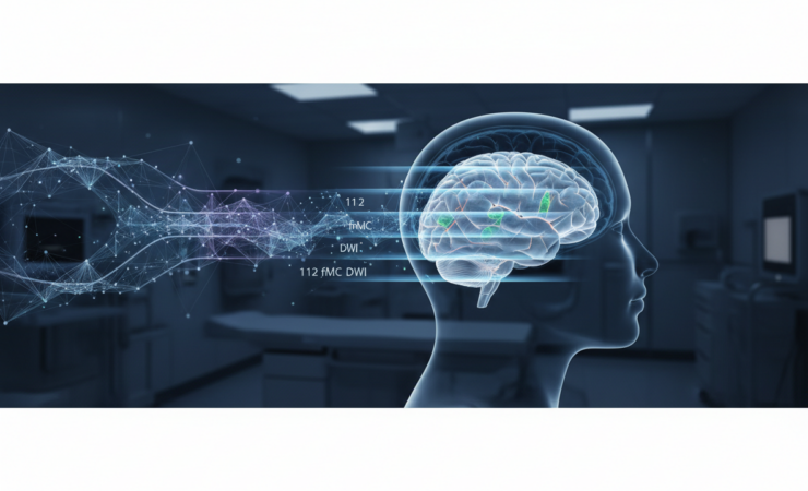
AMF-U-Net uses four separate encoders, one each for T1, T1ce, T2 and FLAIR MRI. This allows the network to extract modality-specific features before combining them. At each encoder scale, a Modality Fusion Module calculates softmax-normalised importance weights and uses those weights to fuse the four streams.
The architecture also incorporates residual connections, which are intended to stabilise optimisation in a deep three-dimensional network. Attention gates are used in the decoder to suppress less relevant activations arriving through skip connections and to focus reconstruction on informative regions and boundaries.
For training, the researchers combined a class-weighted Dice loss with categorical cross-entropy. The two components address different aspects of the task: Dice-based optimisation rewards regional overlap, while class weighting and cross-entropy help reduce the influence of severe voxel-level imbalance.
Two datasets were harmonised before training and evaluation
The study used data from Brain Tumor Segmentation 2023 and the UCSF Preoperative Diffuse Glioma MRI dataset. Because datasets can differ in naming conventions, acquisition characteristics and label definitions, the researchers harmonised the inputs before modelling.
The workflow included modality mapping, spatial normalisation, a source-aware patient-level split and transformation of labels into mutually exclusive classes representing background, necrotic or non-enhancing tumour, oedema and enhancing tumour. Splitting at patient level is important because allowing scans from the same patient to appear in both training and validation data can artificially inflate performance.
Performance was reported for three composite tumour regions commonly used in brain tumour segmentation research: whole tumour, tumour core and enhancing tumour. The Dice coefficient ranges from zero to one, with higher values indicating greater spatial overlap between a predicted segmentation and the reference segmentation.
The strongest overlap was for the whole tumour
On the internal validation cohort, AMF-U-Net achieved a Dice score of 0.845 for whole tumour, 0.813 for tumour core and 0.788 for enhancing tumour. Averaged across those three regions, the macro Dice score was 0.815.
The pattern is informative. Performance was strongest when segmenting the broader whole-tumour region and lower for the more specific enhancing component. That is consistent with the general difficulty of identifying smaller, more heterogeneous structures, although the study’s results alone cannot establish which imaging or biological features caused the difference.
The authors also compared AMF-U-Net with 3D U-Net, nnU-Net, UNETR and Swin UNETR under the same data split. They report that the proposed system performed better on overlap and boundary-distance measures in those comparisons. This same-split approach is valuable because segmentation scores can change materially with different train-validation partitions and preprocessing pipelines.
What the architecture adds
The study’s contribution is not a single new component. Adaptive multimodal fusion, residual connections and attention mechanisms already have established roles in deep learning. The authors instead frame the contribution as their combination within a multi-stream 3D architecture designed around multimodal brain tumour segmentation.
The adaptive fusion mechanism is particularly relevant because it avoids treating the four MRI sequences as if they were equally informative at every layer. In principle, learned weighting allows the network to change the relative contribution of each modality as representations become more abstract.
Attention-guided decoding addresses a different problem. Encoder-decoder networks use skip connections to restore spatial detail that can be lost during downsampling, but those connections can also transmit irrelevant features. Attention gates provide a mechanism for filtering that information before it contributes to the reconstructed segmentation.
Why better segmentation matters
Reliable tumour segmentation can support quantitative imaging research, treatment planning and longitudinal measurement by reducing the amount of manual contouring required. Three-dimensional methods are attractive because they can use spatial context across adjacent slices rather than analysing each image independently.
However, a higher Dice score does not automatically translate into better patient outcomes. Clinical usefulness depends on factors that extend beyond benchmark segmentation accuracy, including performance across scanners and institutions, robustness to missing or degraded sequences, processing requirements, calibration, workflow integration and the consequences of local boundary errors.
The results remain a validation study, not a clinical trial
Several limitations are important when interpreting the findings. The reported 0.815 macro-average Dice score comes from the study’s internal validation cohort. Although two data sources were harmonised, this is not the same as prospective testing in routine clinical practice or independent deployment across hospitals with unseen acquisition protocols.
The study evaluates segmentation performance rather than diagnostic accuracy, treatment decisions or patient outcomes. It therefore cannot show that using AMF-U-Net would improve survival, reduce treatment complications or replace expert radiological assessment.
Comparisons with other neural networks are also conditional on the study’s preprocessing, split and implementation choices. The authors’ same-split comparisons improve fairness within the experiment, but they should not be interpreted as a universal ranking of segmentation architectures across all brain tumour datasets.
Even so, the work illustrates a broader direction in medical imaging AI: rather than simply making networks larger, researchers are increasingly designing architectures around the structure of the clinical data itself. Here, that means recognising that four MRI modalities provide complementary information, that tumour classes are highly imbalanced, and that accurate boundaries require selective recovery of spatial detail.
Source Information
Study: Anushya, A., Almarshdi, R., Alrashidi, B. et al. “AMF-U-Net: an adaptive multimodal fusion residual attention 3D U-Net for boundary-aware brain tumour segmentation.”
Journal: Scientific Reports
Published: 3 October 2026
DOI: 10.1038/s41598-026-72174-x