







Mesenchymal stromal cells are being investigated as treatments for inflammatory disease because they can influence immune responses and support tissue repair. Yet cell preparations are biologically heterogeneous. Even cells grown together under the same conditions can occupy different stages of the cell cycle, raising the possibility that their therapeutic behaviour depends partly on when they are used.
A new peer-reviewed study published in Scientific Reports on 3 October 2026 suggests that deliberately synchronising human umbilical cord mesenchymal stromal cells before treatment can improve their performance in an experimental model of acute colitis. The benefit did not appear to belong to one uniquely superior cell-cycle phase. Instead, the act of synchronising the cell population was associated with better outcomes than using the usual unsynchronised preparation.
Why cell timing could matter
Human umbilical cord mesenchymal stromal cells, or UCMSCs, have attracted interest in regenerative medicine because they are relatively accessible, have low immunogenicity and can release molecules that regulate inflammation. A practical challenge is that cultured cells do not behave as a uniform population. At any moment, individual cells can be resting or progressing through different stages of division.
Yaping Wang and colleagues asked whether controlling that variation could change therapeutic activity. They focused on three broad cell-cycle states: G0/G1, when cells are resting or preparing for DNA replication; S phase, when DNA is copied; and G2/M, when cells prepare for and undergo division.
How the researchers synchronised the cells
The team used serum starvation to concentrate UCMSCs in G0/G1. This produced populations in which approximately 80% to 90% of cells were in that phase. A starvation and double-thymidine-block procedure was used for S-phase synchronisation, yielding about 60% S-phase cells. The researchers then returned S-phase-synchronised cells to complete culture medium for four to eight hours, producing roughly 40% enrichment in G2/M.
They examined whether these manipulations damaged the cells and compared senescence, apoptosis, proliferation, secretion of signalling molecules and immune-regulatory activity across the different preparations. Synchronisation did not significantly increase senescence or apoptosis. G0/G1 cells showed the lowest proliferative potential, while S-phase and G2/M cells proliferated more strongly.
The phases also differed in the molecules they released. S-phase cells secreted less transforming growth factor beta, while prostaglandin E2 secretion showed an increasing trend from G0/G1 through S phase to G2/M. These differences are important because mesenchymal stromal cells are thought to exert much of their therapeutic activity through secreted signals rather than simply replacing damaged tissue.
Testing the cells in acute colitis
The researchers then tested the preparations in mice given 5% dextran sulfate sodium, or DSS, to induce acute colitis. This widely used experimental model damages the intestinal lining and produces inflammation, weight loss and colon shortening. It is useful for comparing biological interventions, but it does not reproduce the full complexity of human inflammatory bowel disease.
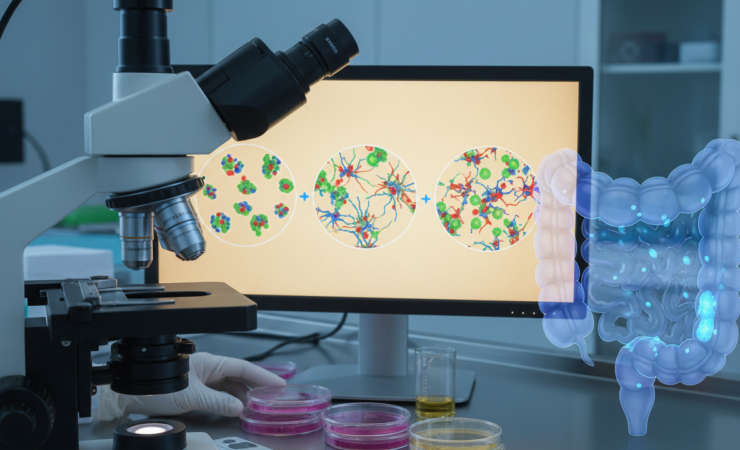
Cells synchronised to different phases were administered on the first and fifth days using tail-vein and intraperitoneal delivery. Disease activity was scored daily. On day seven, the researchers measured colon length and examined colon tissue using histological staining to assess tissue injury, mucus-producing cells and inflammatory infiltration. They also measured regulatory T cells in peripheral blood, spleen and mesenteric lymph nodes.
Synchronised preparations performed better
Compared with unsynchronised UCMSCs, the synchronisation-associated preparations significantly reduced DSS-induced colon shortening, improved histological restoration and reduced inflammatory-cell infiltration. The researchers did not find evidence that one specific cell-cycle phase consistently explained the improvement. Their central conclusion was instead that cell-cycle synchronisation itself enhanced therapeutic efficacy in this acute-colitis model.
The immune findings add nuance to that conclusion. The synchronised cells did not significantly regulate the measured Th1 and Th17 populations, two T-cell groups involved in inflammatory responses. They did, however, show a modulatory effect on regulatory T cells, which help restrain excessive immune activity. This suggests that the observed tissue improvements may involve selective immune pathways rather than a broad suppression of all inflammatory T-cell responses.
A manufacturing question as much as a biological one
The findings point to a potentially useful way of thinking about cell therapies. Researchers often focus on cell source, dose, delivery route and culture conditions. This study suggests that the distribution of cells across the cell cycle may be another property worth controlling before administration.
If the effect proves reproducible, synchronisation could become a pretreatment step used to make therapeutic cell products more consistent. Importantly, the results do not imply that clinicians should select only rapidly dividing cells or only one cell-cycle phase. The experimental advantage was associated with synchronised preparations across phases, which points toward population uniformity or the biological consequences of synchronisation as the more relevant mechanism.
The phase-dependent secretion results also show why this question is complicated. Cells at different stages were not biologically interchangeable. Their proliferation and signalling profiles differed, meaning that synchronisation may alter the composition of signals delivered to inflamed tissue. Establishing which signals are responsible will require more mechanistic work.
What the study cannot yet show
The most important limitation is that the therapeutic experiment was conducted in mice with chemically induced acute colitis. The study does not demonstrate that synchronised UCMSCs improve ulcerative colitis or Crohn’s disease in people, nor does it establish an optimal clinical dose, route or timing schedule.
The degree of synchronisation also varied substantially between phases. G0/G1 preparations reached roughly 80% to 90% synchronisation, S-phase preparations about 60%, and G2/M preparations about 40%. The groups therefore differed not only in cell-cycle identity but also in how homogeneous they were. That makes it harder to isolate exactly why synchronisation was beneficial.
Finally, cell-cycle manipulation adds processing steps to a prospective therapeutic product. Future work would need to establish reproducibility across donors, manufacturing batches and disease models, as well as whether the procedure changes cell persistence, biodistribution or safety after administration.
Why the finding matters
Cell therapies are unusually sensitive to biological variability because the medicine is a living population rather than a chemically identical molecule. The study provides evidence that one source of that variability, cell-cycle state, can be deliberately manipulated without significantly increasing senescence or apoptosis and that doing so can alter treatment performance in vivo.
For regenerative medicine, the broader implication is that manufacturing consistency may influence therapeutic potency in ways that are not obvious from conventional cell identity markers alone. The next step is not to assume that synchronised stem-cell products will work better in patients, but to test whether the effect survives more demanding preclinical models and eventually controlled human studies.
Source Information
Study: Wang, Y., Gao, T., Zhang, X. et al. “Harnessing cell cycle synchronization to boost the therapeutic potential of umbilical cord MSCs against acute colitis.” Scientific Reports (2026).
Published: 3 October 2026
DOI: 10.1038/s41598-026-74352-3