







Laboratory-grown skin models are becoming increasingly sophisticated, but one of their persistent weaknesses is that skin is not simply a stack of epidermal cells. Real human skin contains a dermal matrix, blood vessels, immune cells, hair-associated structures and signalling networks that interact when inflammation develops. Researchers in South Korea have now built a human stem-cell-derived model that brings several of those components together and can reproduce selected features of allergic skin inflammation.
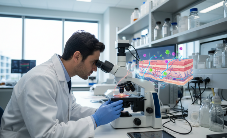
The work, published in Experimental & Molecular Medicine, describes vascularized skin assembloids, or VSAs, created by combining skin organoids derived from human induced pluripotent stem cells with separately generated blood-vessel organoids. The constructs were matured at an air-liquid interface, producing a model with a multilayered epidermis, fibroblast-rich dermal compartment and interconnected endothelial networks with lumen-like structures.
That vascular component matters because inflammatory skin disease involves much more than the outer barrier. Immune cells move through tissue, endothelial cells respond to inflammatory signals and cytokines coordinate responses across different compartments. Conventional skin organoids can reproduce aspects of epidermal organisation, but their lack of vascular structures limits the questions researchers can realistically ask.
Min-Ji Kim, Seunghee Lee and Kyung-Sun Kang therefore used a modular strategy. Instead of trying to coax every tissue component from a single organoid, they generated skin and blood-vessel organoids separately and assembled them under controlled conditions. The resulting VSAs retained epidermal and dermal organisation while adding vascular-like networks and an architecture accessible to experimental exposure from the epidermal side.
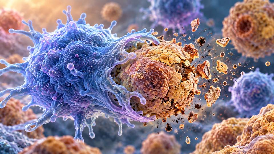
The researchers then tested whether the model could support an immune response. Umbilical-cord-blood-derived mononuclear cells were incorporated into the constructs and the tissue was challenged with inflammatory stimuli. Following lipopolysaccharide stimulation, immune cells accumulated in the upper dermal region and frequently appeared close to CD31-positive vascular structures. In matched non-vascularised skin organoids, infiltrating cells rarely progressed beyond the lower dermis.
Gene-expression measurements reinforced that difference. Vascularised constructs containing immune cells showed stronger induction of endothelial adhesion molecules ICAM1 and VCAM1 and inflammatory cytokines IL-6 and TNF-alpha than comparable non-vascularised constructs. The response suggests that adding vascular architecture changes not only the appearance of the model but also the way inflammatory signalling is organised within it.
The team next exposed the assembloids to house dust mite extract, a clinically relevant allergen associated with atopic dermatitis. The challenge produced several disease-relevant changes, including disruption of barrier-associated features, keratinocyte stress, immune-cell infiltration and activation of type 2 inflammatory mediators. Among the induced signals were IL-4, IL-13, TSLP, CCL17 and CCL26.
Transcriptomic analysis also identified activation of inflammatory, keratinisation and cytokine-signalling pathways. When the researchers compared the response with published transcriptomic data from patients with atopic dermatitis, the stimulated assembloids partially reproduced disease-associated molecular signatures, particularly those involving type 2 and alarmin signalling, keratinocyte stress and programmes related to immune adhesion and infiltration.
A laboratory disease model becomes more useful if it responds plausibly to treatments already known to work in patients. The researchers therefore treated the inflamed assembloids with upadacitinib, a JAK1 inhibitor, and dupilumab, an antibody targeting the IL-4 receptor alpha pathway. Both are established therapies for atopic dermatitis.
Both treatments improved expression of barrier-associated markers including filaggrin, loricrin and claudin-1 while suppressing inflammatory mediators. They also reduced STAT6 phosphorylation and expression of STAT6-associated inflammatory targets. Upadacitinib produced broader transcriptomic changes consistent with suppression of type 2 cytokine networks and modification of epidermal differentiation programmes.
The importance of the result is not that a miniature piece of laboratory skin has recreated atopic dermatitis in full. Rather, the model places epithelial, vascular and immune-associated responses into the same experimentally controllable system. Researchers can determine when components are assembled, when immune cells are introduced, how allergens are applied and how the tissue responds to pharmacological intervention.
That could make the platform useful for studying mechanisms that are difficult to isolate in patients and for screening candidate treatments before more expensive preclinical or clinical work. A human-cell-based model may also complement animal experiments where species differences can complicate translation of inflammatory pathways and drug responses.
There are important limits. The constructs do not reproduce the complete complexity of human skin, chronic disease or circulation in a living body. The vascular networks remain an in vitro approximation, and the current system does not capture the wide patient-to-patient variation seen in atopic dermatitis. The authors specifically identify further vascular maturation, microfluidic integration and patient-specific immune components as areas for development.
The drug experiments should therefore be interpreted as mechanism-consistent validation of the platform rather than evidence that the model can already predict an individual patient’s treatment response. Dupilumab and upadacitinib were selected precisely because their clinical mechanisms are well established, making them useful tests of whether the engineered tissue behaves in a biologically credible way.
Even with those qualifications, the study demonstrates how organoid research is moving beyond reproducing one tissue layer at a time. By combining separately generated components, researchers can build experimental systems in which structural organisation and immune behaviour become increasingly interconnected. For inflammatory skin research, that may provide a more informative bridge between simplified cell cultures and the far greater complexity of human disease.
Source Information
Study Title: Vascularized human skin assembloids model compartmental skin organization and immune responsiveness in vitro
Authors: Min-Ji Kim, Seunghee Lee and Kyung-Sun Kang
Journal: Experimental & Molecular Medicine
Year: 2026
Published: 1 October 2026
DOI: 10.1038/s12276-026-01840-x